
Abstract
The NeuroPlanar Method® is an integrative clinical and performance assessment system leveraging advanced three-dimensional (3D) triplanar motion capture to quantify and correct movement dysfunctions. Utilizing the Kinetisense® platform, this methodology merges biomechanics, neuroanatomy, and artificial intelligence (AI) to deliver precise, individualized interventions for pain management, rehabilitation, and performance optimization. Core components include the KAMS® Functional Movement Screen from Kinetisense®, Functional Planar Mapping® (FPM®), and AI-powered corrective exercise programming. This paper details the biomechanical principles, neurophysiological foundations, technological innovations, and clinical applications of the NeuroPlanar Method®, introducing a new paradigm in human movement science.
1. Introduction
Human movement is a complex interplay between mechanical structures and neural control systems. In real-world tasks, movements occur across three orthogonal planes — sagittal, frontal, and transverse. These planes rarely act in isolation; instead, they interact dynamically to produce functional, coordinated motion.
Traditional biomechanical assessments — single-plane gait observation, isolated joint range-of-motion testing — fail to capture the richness of triplanar integration. Moreover, such assessments often rely on subjective clinician interpretation, which introduces variability and reduces diagnostic accuracy.
The NeuroPlanar Method® was developed to address these limitations by providing a high-resolution, data-driven system that objectively captures and interprets movement across all three planes simultaneously — a "neurological-based" movement screen that provides additional insights alongside other assessments such as FMS® and SFMA®.
2. Triplanar Movement Meets Neuromechanical Intelligence
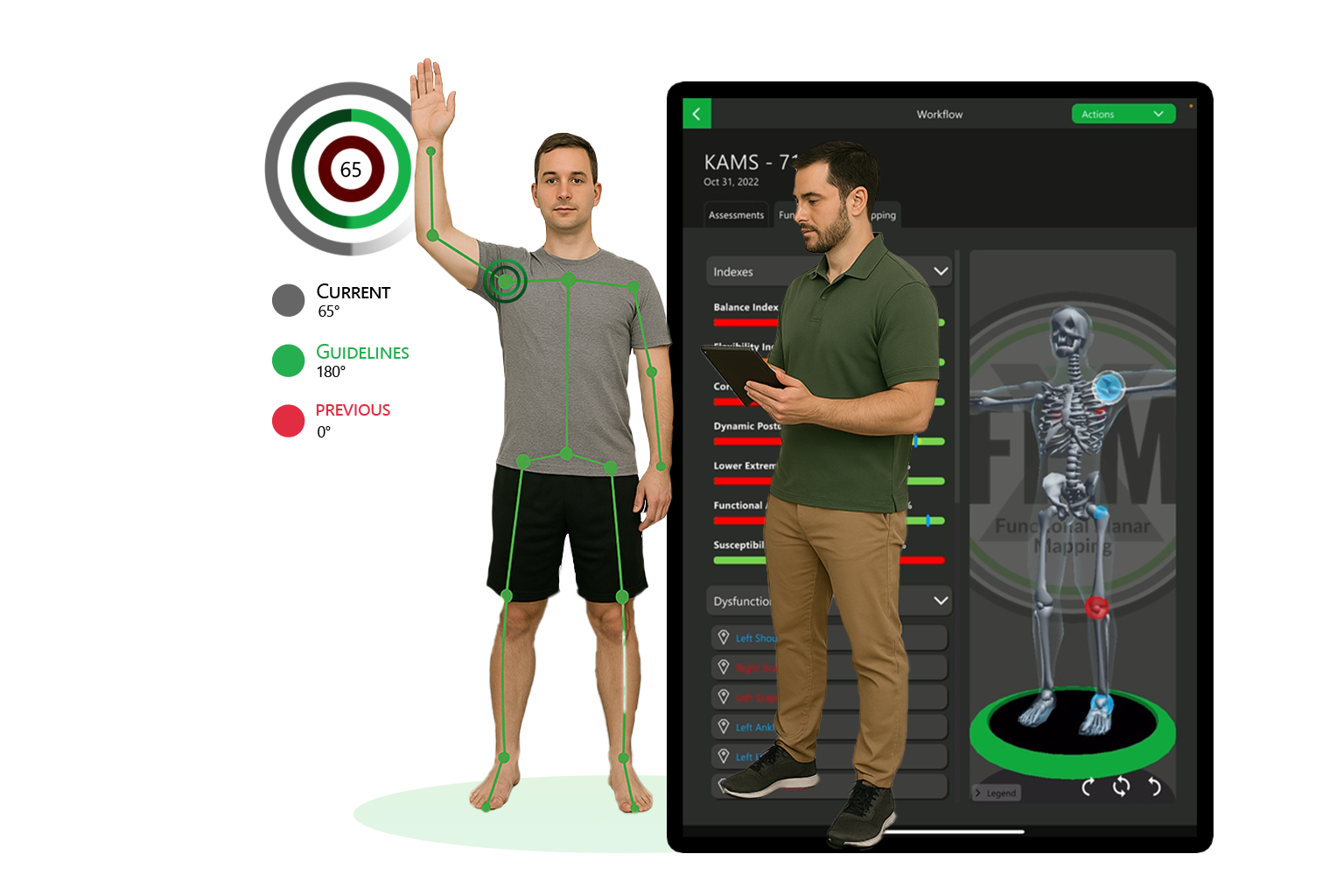
The NeuroPlanar Method® distinguishes itself from conventional tools through its use of the Kinetisense® 3D motion platform. This markerless motion-capture technology measures joint kinematics in real time, delivering precise readings on angles, velocities, and symmetry patterns.
Unlike two-dimensional assessment methods, the platform captures subtle deviations in multiplanar control — such as rotational deficiencies in the transverse plane or lateral instability in the frontal plane — that may be invisible to the naked eye. This approach acknowledges that dysfunction often arises not from absolute weakness or limited mobility, but from inappropriate sequencing, timing, and integration between planes of movement.
2.1 Quantitative and Qualitative Assessment Integration
The NeuroPlanar Method® is unique in its dual approach, capturing both quantitative and qualitative elements of triplanar human movement. Quantitative data, gathered via the Kinetisense® 3D motion platform, includes objective measures such as joint angles, symmetry indices, range-of-motion profiles, velocity patterns, and temporal sequencing. This allows precise benchmarking against normative population values and progress tracking over time.
Qualitative insights are equally critical; through Functional Planar Mapping®, clinicians assess fluidity, coordination, postural control, and movement economy. This combined approach reveals subtle compensations, motor-control deficits, and inefficiencies that may not appear in raw numerical data alone. By integrating both data-driven precision and clinical interpretation, the NeuroPlanar Method® ensures interventions are both scientifically grounded and functionally relevant.
3. Neuroanatomical Basis of Triplanar Control
Movement generation and control are distributed functions, drawing on cortical planning centers, subcortical integration hubs, brainstem reflex loops, and peripheral feedback systems. Triplanar movement demands precise modulation of muscle-recruitment patterns, joint-stabilization strategies, and timing sequences. Errors in these processes can originate from disruptions anywhere along this chain, from cortical planning to peripheral proprioceptor signaling.
The Cerebellum
Refines ongoing motion. It receives proprioceptive input from muscle spindles, Golgi tendon organs, and joint mechanoreceptors, as well as vestibular input for balance. By integrating this sensory information with motor commands from the cortex, the cerebellum adjusts force output and timing, allowing smooth execution of complex movements. This feedforward control predicts and corrects motion before errors occur — vital in sports and rehabilitation contexts where rapid adaptation is required.
The Basal Ganglia
Modulate initiation, scaling, and cessation of movement. In triplanar motion, they ensure that transitions — such as shifting from a sagittal-plane forward lunge to a transverse-plane rotational reach — are fluid and coordinated. Dysfunction in this network, as seen in conditions such as Parkinson's disease, results in rigidity, bradykinesia, and impaired postural reflexes.
The Brainstem
The vestibular nuclei serve as the integration hub for head, neck, and postural alignment in 3D space, synthesizing input from the inner ear, visual system, and somatosensory pathways to maintain balance during multiplanar transitions. In the NeuroPlanar Method®, deficits in vestibular integration often present as delayed corrective responses, excessive sway, or asymmetric weight distribution.
The Motor Cortex
Including premotor and supplementary motor areas (SMA), provides high-level programming for coordinated, voluntary multiplanar tasks. The SMA contributes to internally generated movements and complex sequences, while the premotor cortex is more involved in externally cued motion. Both regions rely on continuous feedback from proprioceptive and visual systems to adapt plans in real time.
The Thalamus
Acts as a central relay station, transmitting sensory feedback to cortical areas for conscious and subconscious adjustment. Its role in proprioceptive processing is crucial for aligning intended motion with actual performance, especially when movement involves rapid directional changes.
3.1 Myelination Patterns, Neuroplasticity, and Movement Efficiency
Myelination — the process of insulating axons with myelin sheaths — improves conduction velocity and fidelity of neural signals. In multiplanar movement, faster conduction enables precise timing between sensory feedback and motor execution. Myelination progresses from primary motor and sensory areas in infancy to associative cortical regions in adolescence, paralleling the development of more complex movement skills. In adults, high-level performance correlates with optimized, reinforced myelinated pathways, while injury or disuse can lead to conduction delays.
The NeuroPlanar Method® leverages targeted triplanar repetition to stimulate adaptive myelination and reinforce efficient motor patterns. This approach aligns with neuroplastic principles, allowing clinicians to retrain lost or underdeveloped pathways.
3.2 Planar Plasticity: Adaptive Control Across Three Planes
Planar plasticity describes the nervous system's ability to efficiently integrate and transition between sagittal, frontal, and transverse plane movements. This capacity is critical for performance and injury prevention. High planar plasticity results in seamless load transfer, optimal energy efficiency, and reduced stress on passive structures. Low planar plasticity leads to asymmetrical loading, delayed stabilization, and greater injury susceptibility.
The NeuroPlanar Method® quantifies planar plasticity using Functional Planar Mapping® to identify deficits, and prescribes interventions — ranging from proprioceptive drills to strength and mobility training — that restore adaptive capacity.
4. Core Methodology Components
KAMS® Functional Movement Screen
Objective, population-referenced 3D movement screening from Kinetisense®.
Functional Planar Mapping® (FPM®)
Real-time stability and mobility mapping across all planes.
AI-Powered Corrective Exercise Programming
Automated, personalized exercise plans.
Integrated Therapy & Training
Soft-tissue therapy (e.g. Active Release Technique®), kinesiology taping (e.g. RockTape®), joint mobilization, and neuromuscular re-education, integrated with strengthening and sport-specific training.
5. Clinical & Performance Applications
6. Discussion
The NeuroPlanar Method® integrates biomechanics, neuroscience, and AI-driven corrective intervention to deliver scalable, evidence-based care — bridging objective measurement and clinical judgment in a single, repeatable workflow.
7. Conclusion
The NeuroPlanar Method® provides a comprehensive, neurophysiologically grounded approach to movement assessment and optimization — a new paradigm in human movement science.
References
- Manto, M., & Huisman, T. A. (2016). The Cerebellum: Disorders and Treatment. Cambridge University Press.
- Bastian, A. J. (2006). Learning to predict the future: The cerebellum adapts feedforward movement control. Current Opinion in Neurobiology, 16(6), 645–649.
- Fields, R. D. (2008). White matter in learning, cognition and psychiatric disorders. Trends in Neurosciences, 31(7), 361–370.
- Zatorre, R. J., Fields, R. D., & Johansen-Berg, H. (2012). Plasticity in gray and white: Neuroimaging changes in brain structure during learning. Nature Neuroscience, 15(4), 528–536.
- Dietrichs, E., & Haines, D. E. (2002). Interconnections between the cerebellum and basal ganglia. Journal of Anatomy, 199(5), 539–551.
- Nachev, P., Kennard, C., & Husain, M. (2008). Functional role of the supplementary and pre-supplementary motor areas. Nature Reviews Neuroscience, 9(11), 856–869.
- Sherman, S. M., & Guillery, R. W. (2002). The role of the thalamus in the flow of information to the cortex. Philosophical Transactions of the Royal Society B, 357(1428), 1695–1708.
- Taube, W., et al. (2007). Brain plasticity and sensorimotor learning in balance control. Gait & Posture, 26(4), 650–659.
- Peterson, D. S., & Horak, F. B. (2016). Neural control of walking in people with Parkinsonism. Physiology, 31(2), 95–107.